
Datei:a-Image-obtained-with-a-microscope-of-the-micro-needle-distribution-in-Ferrocell-for-a W640.jpg


Größe dieser Vorschau: 832 × 966 Pixel.
Originaldatei (832 × 966 Pixel, Dateigröße: 328 KB, MIME-Typ: image/jpeg)
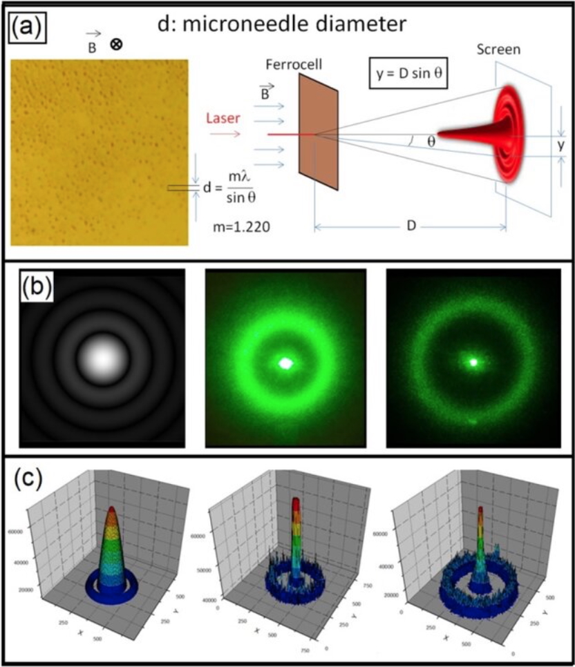
(a) Image obtained with a microscope of the micro-needle distribution in Ferrocell for a magnetic field perpendicular to the plane of the plate together with the diagram for the Airy pattern obtained by diffracting a 532 nm green laser. With this apparatus, we can measure the diameter of micro-needles. (b) Theoretical and experimental Airy patterns obtained with Ferrocell with an external magnetic field of 400 G and with a field of 600 G. The average diameter d of the micro-needles is around 4.3 μm. In (c), we have the intensity graph and these plots were obtained transforming the images in grayscale.
Dateiversionen
Klicke auf einen Zeitpunkt, um diese Version zu laden.
| Version vom | Vorschaubild | Maße | Benutzer | Kommentar | |
|---|---|---|---|---|---|
| aktuell | 08:25, 11. Mär. 2026 | | 832 × 966 (328 KB) | Nikolaos (Diskussion | Beiträge) | |
| 08:24, 11. Mär. 2026 | 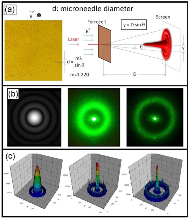 | 640 × 742 (78 KB) | Nikolaos (Diskussion | Beiträge) | (a) Image obtained with a microscope of the micro-needle distribution in Ferrocell for a magnetic field perpendicular to the plane of the plate together with the diagram for the Airy pattern obtained by diffracting a 532 nm green laser. With this apparatus, we can measure the diameter of micro-needles. (b) Theoretical and experimental Airy patterns obtained with Ferrocell with an external magnetic field of 400 G and with a field of 600 G. The average diameter d of the micro-needles is around 4.… |
Du kannst diese Datei nicht überschreiben.
Dateiverwendung
Die folgende Seite verwendet diese Datei:
